Hooke’s Micrographia Reveals a World Under the Lens
Detailed drawings of magnified insects and materials gave the word cell to biology.
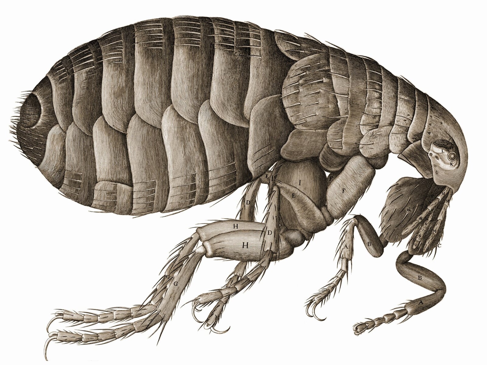
In 1665, Robert Hooke, Curator of Experiments at the Royal Society of London, published Micrographia, an illustrated book of observations. Utilizing a compound microscope of his own design, Hooke examined a wide range of common objects and small organisms under magnification, transcribing what he saw into elaborate drawings. His artistic training, which had begun in his youth as an apprentice in London, enabled him to render minute physical textures with exceptional clarity. Micrographia offered contemporaries detailed visual depictions of structures invisible to the unassisted eye, encouraging broader scientific investigation into the microscopic domain.
Among the numerous subjects Hooke inspected was the structure of common cork. Observing the porous framework of the material under the lens, he coined the word cell to name the minuscule compartments he noted within the sample. This terminology subsequently became fundamental to modern biology. Hooke did not restrict his study to organic tissues; Micrographia also presented examinations of non-living materials and stellar phenomena. The volume featured illustrations of the Pleiades star cluster alongside depictions of lunar craters, whose shapes led Hooke to argue that the Moon probably had a gravitating principle like Earth's.

Keep reading in Yester
Coming soon to the App Store
Keep following the story
Sources
- Robert Hooke (MacTutor, University of St Andrews)
- Robert Hooke (1635-1703) (University of California Museum of Paleontology)
- Robert Hooke (Encyclopaedia Britannica)
- Micrographia by Robert Hooke (Project Gutenberg)
- The Monument to the Great Fire of London (London Museum)
- Print of Bethlem Hospital, collection object P_1977-U-824 (British Museum)
- Robert Hooke
Image: Robert Hooke