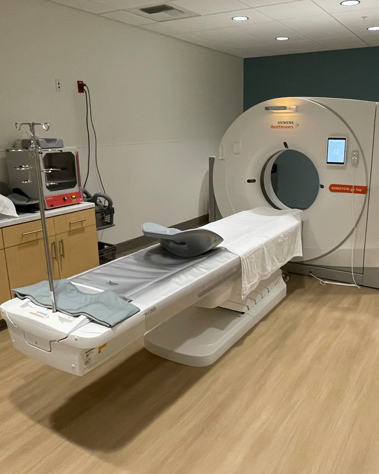
The First CT Scan Looks Inside a Patient’s Head
A computer turned many X-ray views into a slice through the body.
Between 1967 and 1972, British electrical engineer Godfrey Hounsfield developed the first commercially viable computed tomography scanner at the research laboratories of EMI. His work built on mathematical principles that extended back to the 1917 Radon transform, alongside earlier concepts such as William H. Oldendorf's 1963 patent for investigating interior objects obscured by dense materials. Hounsfield's innovative device combined diagnostic physics with digital calculation, earning him the 1979 Nobel Prize in Physiology or Medicine alongside South African-American physicist Allan MacLeod Cormack.
The first clinical test came at Atkinson Morley's Hospital in Wimbledon, south London, where consultant radiologist Jamie Ambrose agreed to work with Hounsfield and a prototype EMI brain scanner was installed. On 1 October 1971 the team scanned its first patient, a woman in her early forties with a suspected brain tumor. The process was slow: the scan took about 30 minutes, and the data then had to be driven across town on magnetic tape and processed for hours before an image appeared. It showed a cystic mass in the frontal lobe. When Ambrose presented the first clinical images to the British Institute of Radiology on 20 April 1972, they helped dispel radiologists' doubts about the technique.

Keep reading in Yester
Coming soon to the App Store
Keep following the story
Sources
- Godfrey N. Hounsfield, Biographical (NobelPrize.org)
- Allan M. Cormack, Biographical (NobelPrize.org)
- Perspectives: With a little help from my friends (NobelPrize.org)
- Fifty Years Ago, the First CT Scan Let Doctors See Inside a Living Skull (Smithsonian Magazine)
- How This Record Company Engineer Invented the CT Scanner (IEEE Spectrum)
- What are the Radiation Risks from CT? (U.S. Food and Drug Administration)
- Computed Tomography (CT) (U.S. Food and Drug Administration)
- CT scan
Image: USDAgov